
Input and output nerves meet at
- (a)liver
- (b)central nervous system
- (c)heart
- (d)None of the above
Answer
Why
Correct: B, central nervous system. Translate the stem's everyday words into the textbook ones and the answer is almost a definition. The 'input' nerves are the sensory or afferent fibres, which carry information inward from receptors (in the skin, eye, ear, tongue, nose and the stretch receptors of muscle) towards the centre. The 'output' nerves are the motor or efferent fibres, which carry instructions outward from the centre to the effectors, meaning skeletal muscles and glands. The place where the incoming stream is read and the outgoing stream is issued is by definition the integrating centre, and in a vertebrate that is the central nervous system: the brain and the spinal cord together. The anatomy is unusually literal about this. Afferent and efferent fibres travel side by side in a mixed spinal nerve out in the limb, but they part company the moment they reach the cord: sensory fibres enter through the dorsal, or posterior, root, motor fibres leave through the ventral, or anterior, root, a separation known as the Bell–Magendie law. The two therefore physically converge inside the grey matter of the spinal cord and nowhere in the periphery. The clearest working example is the reflex arc that NCERT uses: touch something hot and the chain runs receptor → sensory neuron → dorsal root → relay neuron in the grey matter of the cord → motor neuron → ventral root → muscle of the arm. The hand is pulled back before the brain has registered pain, because the connection is made in the cord and only afterwards does the information travel up to the brain. That short circuit is the whole reason reflexes are fast. And the junction itself is not a fusion but a synapse: a gap of a few tens of nanometres across which the electrical impulse is converted into a chemical signal by a neurotransmitter such as acetylcholine, then converted back. Because transmitter is released on one side and detected on the other, the synapse acts as a one-way valve and fixes the direction of travel through the whole arc.
Why the others are wrong
- (a)liver: The largest gland in the human body and its principal chemical factory: it secretes bile, stores glucose as glycogen, deaminates surplus amino acids and builds urea, detoxifies drugs and alcohol, and makes most of the plasma proteins. None of that is integration of nervous signals. The liver does have a nerve supply, autonomic fibres reaching it through the hepatic plexus, but that traffic is output only: the nervous system instructs the liver, and no sensory pathway terminates there to be handed on to a motor one. It is an effector organ, and this option is on the list mainly because it is the organ candidates associate with 'does many things at once'.
- (c)heart: The genuinely tempting option, because more is true of the heart than of the liver here. The heart does receive both arms of the autonomic nervous system (sympathetic fibres that raise rate and force, and the parasympathetic vagus that slows it) so two nerve supplies really do arrive there. It also has its own conducting system: the sino-atrial node, the atrioventricular node, the bundle of His and the Purkinje fibres, which make the heart myogenic, able to beat with every nerve to it cut. But that conducting tissue is specialised cardiac muscle, not nerve, and the autonomic supply is purely efferent: it carries orders in, not information out. Stretch and pressure receptors in the heart and the great vessels send their signals away to the cardiovascular centre in the medulla oblongata, and the corrective instruction is issued from there. The loop closes in the brain stem, not in the chest.
- (d)None of the above: Not available, because one of the three preceding options is exactly right. A candidate may be tempted here by a half-remembered fact (that some reflexes never reach the brain) and conclude that no single named structure can be the meeting point. That reasoning defeats itself: the spinal cord where those reflexes are completed is part of the central nervous system, so 'CNS' covers the reflex case and the deliberate case alike, which is precisely why it is the safe answer rather than 'brain'.
Concept
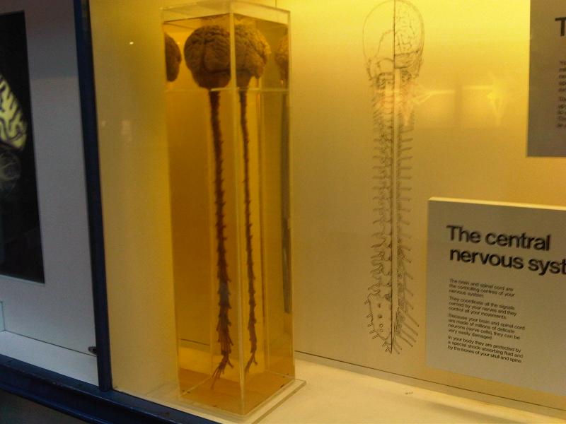
The human nervous system is built in two halves that divide the labour of control. The central nervous system (the brain and the spinal cord) receives, sorts and interprets information and decides what to do about it. The peripheral nervous system is the wiring that connects it to everything else: twelve pairs of cranial nerves leaving the brain and thirty-one pairs of spinal nerves leaving the cord, each carrying afferent fibres in and efferent fibres out. The efferent half of the peripheral system splits again into the somatic division, which drives skeletal muscle under voluntary control, and the autonomic division, which runs the viscera involuntarily and is itself divided into the sympathetic system of fight-or-flight and the parasympathetic system of rest-and-digest. The functional unit throughout is the neuron (a cell body with branching dendrites that receive signals and a single long axon that transmits them) and neurons never touch. They communicate across synapses, where a neurotransmitter carries the message chemically from one cell to the next in one direction only. Stack these facts and the architecture of every response becomes the same three-step sequence: receptor, integrating centre, effector. Only the middle step is negotiable, and it always lies within the central nervous system.
Two moves solve this question. First, translate: 'input nerve' means sensory or afferent, 'output nerve' means motor or efferent, and once those words are on the page the question is asking where a sensory neuron hands over to a motor neuron. Second, test each option for whether both kinds of fibre are present. The liver fails immediately, since it receives autonomic instructions and returns no sensory pathway for handover. The heart is the trap and deserves a moment, because it does receive two nerve supplies and does have an internal conducting network, but sympathetic and vagal fibres are both output, and the conducting system is modified muscle rather than nerve, so no afferent-to-efferent junction exists there either. Only the central nervous system has both streams under one roof, and the anatomy makes the meeting point literal: sensory fibres enter the cord by the dorsal root and motor fibres leave by the ventral root, so the crossover happens in the grey matter. One honest refinement belongs on this card for the student who reads further. The gut wall carries an enteric nervous system, sometimes called the second brain, which does contain its own sensory neurons, interneurons and motor neurons and can run local reflexes without consulting the brain or cord at all. That is a real exception to the rule that all integration is central, but it sits in the wall of the intestine, not in the liver and not in the heart, so it rescues no option on this list.
Key facts
- The central nervous system consists of the brain and the spinal cord. The peripheral nervous system consists of 12 pairs of cranial nerves and 31 pairs of spinal nerves (8 cervical, 12 thoracic, 5 lumbar, 5 sacral and 1 coccygeal) and its efferent side divides into the somatic and the autonomic, the autonomic dividing again into sympathetic and parasympathetic.
- By the Bell–Magendie law, sensory fibres enter the spinal cord through the dorsal root and motor fibres leave through the ventral root, so the two streams physically converge in the grey matter of the cord and not anywhere in the periphery.
- The reflex arc runs receptor → sensory neuron → interneuron in the spinal cord → motor neuron → effector. The knee-jerk stretch reflex is the simplest of all, monosynaptic, with the sensory neuron synapsing straight on to the motor neuron and no interneuron in between; the withdrawal reflex from a hot object uses interneurons and is complete before the brain registers pain.
- Neurons never touch. They meet at synapses, gaps of a few tens of nanometres bridged by a chemical transmitter such as acetylcholine, which is released on the pre-synaptic side only: making the synapse a one-way valve and fixing the direction of flow through the arc.
- The adult human brain weighs roughly 1.3 to 1.4 kg and contains of the order of 86 billion neurons. The spinal cord is only about 43 to 45 cm long, ending near the first or second lumbar vertebra, below which the nerve roots continue downward as the cauda equina, which is why a lumbar puncture is taken below that level.
Study next
Common traps
- Answering 'brain' where the option list offers 'central nervous system': many reflex handovers occur in the spinal cord, so only the wider term is safe
- Choosing the heart because it receives sympathetic and vagal fibres and has its own conducting system; both nerve supplies are output, and the conducting tissue is cardiac muscle rather than nerve
- Confusing the ganglia of the autonomic system with integration centres: the synapse there is between two motor neurons, preganglionic and postganglionic, with no sensory fibre involved
BPSC put this in plain language rather than in textbook vocabulary: 'input and output nerves' instead of afferent and efferent, so the difficulty is one of translation, not of biology, and the option list sets one nervous structure against two unrelated organs, which is the Commission's standard way of building a science distractor set on this paper. The 71st CCE in 2025 used the same one-line shape on the body's other coordination system, asking which class of gland secretes hormones. UPSC goes a level deeper into the same anatomy: it names a specific structure and asks what it controls, as with the medulla oblongata and the regulation of swallowing and vomiting in 2007, or asks which gland escapes the control of the master gland, as with the parathyroid in 1997.
Related PYQs
Which one of the following parts of the human brain is the regulating centre for swallowing and vomiting?
- (a) Cerebellum
- (b) Cerebrum
- (c) Medulla oblongata
- (d) Pons
Answer(c) Medulla oblongata
The same principle at higher resolution. Swallowing and vomiting are reflexes whose sensory input and motor output are joined in the medulla oblongata: a part of the brain stem, and therefore of the central nervous system. UPSC asks which room in the building; BPSC asks which building.
The pituitary gland by virtue of its tropic hormones controls the secretory activity of other endocrine glands. Which one of the following endocrine glands can function independent of the pituitary gland?
- (a) Thyroid
- (b) Gonads
- (c) Adrenals
- (d) Parathyroid
Answer(d) Parathyroid
The identical question: where does control sit? asked of the body's other coordination system. The pituitary is the endocrine integrating centre and is itself directed by the hypothalamus, a part of the brain, which is the exact point at which nervous and hormonal control join hands; the parathyroid is the exception because it responds straight to blood calcium.
Hormones are secreted by :
- (a) Sebaceous glands
- (b) Reproductive glands
- (c) Exocrine glands
- (d) Endocrine glands
Answer(d) Endocrine glands
BPSC returned to control and coordination on the 71st CCE with the same one-line design, this time on the chemical arm rather than the nervous one. Both questions reward the candidate who holds the general category (endocrine glands, central nervous system) rather than a single organ, since the narrower answer is what the distractors are made of.
Practice
- practice, not a real PYQ
In a spinal nerve, the sensory fibres enter the spinal cord through the
- (a)ventral root
- (b)dorsal root
- (c)cauda equina
- (d)grey commissure
Answer(b) dorsal root: under the Bell–Magendie law, afferent or sensory fibres enter the cord by the dorsal (posterior) root while efferent or motor fibres leave by the ventral (anterior) root. The cauda equina is the bundle of nerve roots continuing below the end of the cord, and the grey commissure is the bridge of grey matter around the central canal. - practice, not a real PYQ
Which one of the following is NOT a part of the central nervous system?
- (a)Cerebellum
- (b)Medulla oblongata
- (c)Spinal cord
- (d)Vagus nerve
Answer(d) Vagus nerve: it is the tenth cranial nerve and therefore part of the peripheral nervous system, carrying the parasympathetic supply to the heart, lungs and gut. The cerebellum and medulla oblongata are regions of the brain, and the spinal cord is the other half of the central nervous system.